Planning radiotherapy for eye cancer
The radiotherapy team plan your radiotherapy before you start treatment. This means working out the dose of radiotherapy you need and exactly where you need it. The plan they create is just for you.
Planning treatment
During your treatment, you will meet a number of people. They work together to plan and give you your treatment. These people include:
-
a radiotherapy doctor - a doctor who specialises in treating cancer with radiotherapy
-
a radiographer - a person trained to give radiotherapy treatment and operate the machines
There are different ways of having radiotherapy to the eye:
-
external beam therapy - for this treatment, a machine directs radiotherapy beams at the cancer from outside the eye
-
internal radiotherapy (brachytherapy) - a small radioactive disc is stitched to the eye. This gives a high dose of radiation to the eye cancer
External radiotherapy
You may have one of the following types of external radiotherapy for eye cancer:
-
stereotactic radiosurgery
-
proton therapy
Stereotactic radiosurgery
Stereotactic radiosurgery uses several small thin radiation beams. They target a large dose of radiotherapy on the cancer. Although it’s called radiosurgery, no surgery is involved.
Several types of machines deliver this treatment:
- Linac (linear accelerator)
- Gamma Knife
- CyberKnife
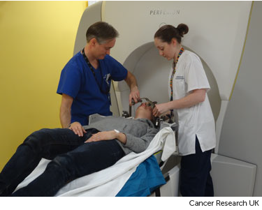
Your head needs to be kept as still as possible during stereotactic radiotherapy. This is called immobilisation. To do this you might have:
- a mask
- a head frame
Proton therapy
Proton therapy machines treat eye cancers using a type of radiation known as proton radiation. The proton beams are aimed precisely at the cancer.
Before proton therapy you have an operation to put tiny metal clips (tags) on the outer wall of the eye. You cannot see them, but they are visible on scans. This is important as they help your radiation doctors plan your treatment. You usually have the clips put in under general anaesthetic. They normally stay in place after treatment. They will not affect your eyesight.
The surgery in preparation for the proton therapy is usually done at your ocular oncology centre, but you will have proton therapy at the Clatterbridge Cancer Centre near Liverpool.
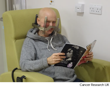
You have all your planning and treatment while sitting in a chair. It is designed for this type of radiotherapy. You have 1-2 planning appointments. They involve having:
- a mask made
- a mouthpiece made that attaches to a frame
- having x-rays
The mask and frame make sure:
- you keep your head completely still during planning and treatment
- you have radiotherapy to the exact same place for each treatment
The radiation doctors ensure they have all the information from the x-rays to plan your treatment. You wait in the department while this is being done. Sometimes, they may need more x-rays. All together, this can take up to 3 hours. Afterwards you may have an appointment to attend a second session.
Between appointments, the radiation doctors work out your individual radiotherapy treatment.
At your second appointment, the doctors check your treatment plan is accurate and may make minor changes. This appointment takes about 30 minutes.
Internal radiotherapy
Before you start the treatment, you have an appointment to plan your radiotherapy. Your radiation oncologist works with other radiation specialists to calculate the treatment dose. The amount you have and the time it is in place depends on:
-
the thickness of the cancer
-
the size of the cancer
-
the size of the plaque used
Your specialist works out the correct dose for you. Your radiotherapy team will let you know when your treatment will start.
Radiotherapy mask (mould)
A radiotherapy mask is also called a shell. It keeps your head still each time you have your radiotherapy. So your treatment is as accurate as possible.
You can see through most types of masks, as they usually have lots of small holes. The radiographers might make marks on them. They use the marks to accurately line up the radiotherapy machine for each treatment.
Your team might make the mask in the mould room of the radiotherapy department or during your CT planning session. It takes between 10 to 45 minutes depending on the type of mask.
Before making the mask
The mask is normally made directly against your skin. It's helpful to wear clothing that you can easily take off from around your neck. You also need to take off any jewellery from that area.
Having a lot of facial hair can make it difficult to make a head mask. The radiotherapy staff will tell you about any hair issues at your planning session.
Making the mask
A mould technician or radiographer uses a special kind of plastic that they heat in warm water. This makes it soft and pliable. They put the plastic on to your face so that it moulds exactly. It feels a little like a warm flannel and is a mesh with holes in so you can breathe.
After a few minutes the mesh gets hard. The technician takes the mask off and it is ready to use.
The mask is kept in the radiotherapy department and you wear it for each treatment. Or you might have a mask made for a single treatment.
The video below shows what happens when you have your mesh mask made. The video is about 1 and a half minutes long.
Voiceover: Making a mesh mask for radiotherapy takes a few minutes.
Radiographer: I am just going to heat this up now if you just keep nice and still there and just want to close your eyes for us.
Voiceover: The radiographer softens the mask by putting it in warm water for a minute or two. When the radiographer puts the mask on to your face it will feel warm and damp. They then clip it to the bed that you are lying on. It takes a minute or two to dry into the shape of your face. The radiographers will mark the mask where the light lines are.
Radiographer: Okay, you are just going to feel us pressing down on the mask there; you are doing really well are you still okay?
Patient: Mmm...
Voiceover: They use the marks on the mask to line up the machine each time you have treatment. The mask keeps you head still and makes sure that your treatment is directed at the cancer. They put your name on the mask and keep it in the radiotherapy department ready for your treatment.
Patient: They um told me about the procedure, a mask being fitted, uumm that it would be moulded to the shape of my face. Umm which they did, three lovely girls umm put my mind at ease, sat me down, heated the mask, moulded it around my face, um not an uncomfortable thing at all to go through.
Head frame
Your radiotherapy team might fit a head frame if you are going to have treatment from a GammaKnife machine. They usually fit the frame on the same day as your radiotherapy treatment.
They attach the head frame to your skull using 4 pins. Before they attach it, you have 4 injections of local anaesthetic at the points where the frame attaches to your head. This takes about 10 minutes. The head frame is then fixed into the radiotherapy machine while you are lying on the treatment couch.
As they fit the frame, you feel some pressure and tightness, but it usually feels better within a few minutes. You then have your radiotherapy treatment on the same day.


You usually have a head frame fitted for a single treatment or a small number of treatments. If you are going to have a number of treatments, you have the frame fitted and removed each time.
After your planning session
You might have to wait a few days or up to 3 weeks before you start treatment.
During this time, the physicists and your radiotherapy doctor (clinical oncologist) will decide the final details of your radiotherapy plan. They make sure that the area of the cancer will receive a high dose and nearby areas receive a low dose. This reduces the side effects you might get during and after treatment.


