Having external radiotherapy for eye cancer
External radiotherapy means having radiation from a machine outside the eye. It only treats the area of the body the radiation is directed at.
When you might have it
You may have external radiotherapy on its own. Or you might have it with other treatments such as surgery and chemotherapy. The treatment you have and the order you have it depends on the type and size of the cancer. The types of eye cancers that are treated with external beam radiotherapy include:
Eye melanoma
Radiotherapy is often used to treat melanoma of the eye. Internal radiotherapy (brachytherapy) is usually used for small or medium sized eye melanomas. If internal radiotherapy is not suitable for your cancer, you might have external radiotherapy.
You may have surgery before radiotherapy. This depends on the size of the cancer and where it is. Having more than one type of treatment could mean that you can keep some, or all of your sight.
Eye lymphoma
You usually have external beam radiotherapy for lymphoma of the eye.
Your doctor may suggest radiotherapy treatment similar to that used for non-Hodgkin lymphoma. Radiotherapy for eye lymphoma can include both eyes. This is because there is a high chance of the cancer being present in both eyes.
Some eye lymphomas can spread to the brain. In this situation you have radiotherapy to the eyes and brain. It can clear the cancer in the eye and also help to stop it coming back in the brain or spinal cord.
You may have chemotherapy as well as radiotherapy.
Lacrimal gland cancer
You might have external beam radiotherapy following an operation to remove your lacrimal gland cancer. This aims to treat any cancer cells left behind and reduce the risk of it coming back.
Sometimes it is possible to remove the lacrimal gland cancer, without removing the whole eye.
Squamous cell skin cancer
You may have external radiotherapy on its own for squamous cell skin cancer of the eyelid when surgery is too risky. Or you might have radiotherapy after surgery in one of the following situations:
- your cancer has spread to lymph nodes or nerves
- there is a high risk that it may come back
Types of external radiotherapy
There are different types of machines that give external radiotherapy. Some are better for treating cancers on the surface of the eye. Others are best for eye cancers that are deeper in the socket. Your radiotherapy specialist (clinical oncologist) carefully chooses the machine that is right for your type of eye cancer.
Types of external radiotherapy you may have for eye cancer are:
- stereotactic radiosurgery
- proton therapy
Stereotactic radiosurgery
Stereotactic radiosurgery uses lots of small thin radiation beams. They target a large dose of radiotherapy on the cancer. You may have a single treatment, or up to 5 treatments.
As treatment is targeted at the cancer, there is less radiation to healthy tissue and fewer side effects. Although it’s called radiosurgery there is no surgery involved.
There are several types of machines that deliver this treatment:
- Linac (linear accelerator)
- Gamma Knife
- CyberKnife
Proton therapy
Proton therapy machines treat eye cancers using a type of radiation known as proton radiation. The proton beams are aimed precisely at the cancer. This type of radiation reduces the risk of damage to the surrounding healthy eye tissue.
You usually have treatment every day for 4 to 5 days.
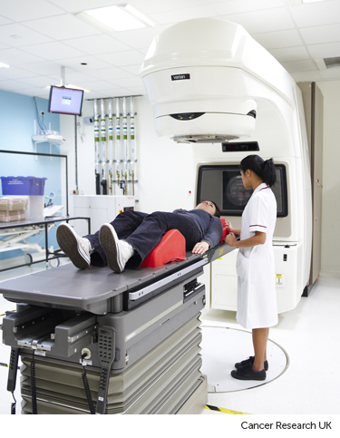
The radiotherapy room
Radiotherapy machines are very big and could make you feel nervous when you see them for the first time. The machine might be fixed in one position. Or it might rotate around your body to give treatment from different directions. The machine doesn't touch you at any point.
Before your first treatment, your  will explain what you will see and hear. In some departments, the treatment rooms have docks for you to plug in music players. So you can listen to your own music while you have treatment.

During treatment
Stereotactic radiosurgery
On the day of your treatment, your radiographer takes you to the treatment room. When you are lying comfortably on the couch your head frame or mask is fitted. The radiographers leave the room before treatment starts. Although you are alone, they can see you from the control room.
Once everything is in place, the couch slides into the machine and the treatment begins. You are awake the whole time.
You won't feel anything while you are having treatment. It can take a few hours. Before you start your radiographer will tell you exactly how long your treatment takes.
Proton therapy
You might have local anaesthetic eye drops just before your treatment. This helps reduce the amount of eye movement. Each treatment takes about 30 seconds, but the appointment takes around 30 minutes. In this time the radiographer makes sure:
-
you are in the correct position
-
you have the correct dose to the right part of the eye
You are in the room on your own during treatment, and you don’t feel or see anything. Sometimes you can hear a hum or buzz while you have it.
After each appointment, a pad is placed over your eye to protect it until the anaesthetic wears off.
You won't be radioactive
External radiotherapy does not make you radioactive. It's safe to be with other people throughout your course of treatment, including pregnant women and children.
Travelling to radiotherapy appointments
You might have to travel a long way each day for your radiotherapy. This depends on where your nearest cancer centre is. This can make you very tired, especially if you have side effects from the treatment.
You can ask your radiographers for an appointment time to suit you. They will do their best, but some departments might be very busy. Some radiotherapy departments are open from 7 am till 9 pm.
Car parking can be difficult at hospitals. Ask the radiotherapy staff if you are able to get free parking or discounted parking. They may be able to give you tips on free places to park nearby.
Hospital transport may be available if you have no other way to get to the hospital. But it might not always be at convenient times. It is usually for people who struggle to use public transport or have any other illnesses or disabilities. You might need to arrange hospital transport yourself.
Some people are able to claim back a refund for healthcare travel costs. This is based on the type of appointment and whether you claim certain benefits. Ask the radiotherapy staff for more information about this and hospital transport.
Some hospitals have their own drivers and local charities might offer hospital transport. So do ask if any help is available in your area.
Side effects of treatment
Radiotherapy for eye cancer has some side effects. The side effects you might have depend on which part of your eye has been treated.


