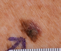
Pictures of abnormal moles, melanoma and skin changes
The pictures on this page are of abnormal moles or areas of skin that:
- are melanoma
- may appear to be melanoma, but were found to be non cancerous (
benign) 
Most of these pictures show what the mole or skin changes look like close up.
The pictures below have been provided by the St John’s Institute of Dermatology at Guy’s and St Thomas’ Hospital.
These pictures are just a guide. Without further tests, it's not possible to work out what is a melanoma or not. If you’re worried about any moles or skin changes, it is always important to get them checked by your GP.
Melanoma that has developed from a suspicious dark mole
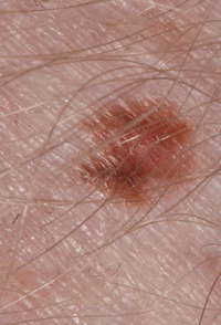
Suspicious irritated mole found not to be melanoma
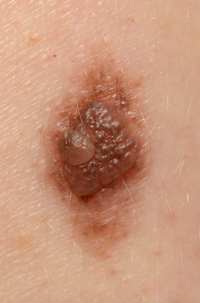
Melanoma from a mole that was once an even colour and shape but has now changed
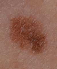
Melanoma from a mole with changing shape and colour
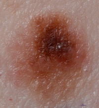
Melanoma that has developed from a long standing mole that is starting to spread
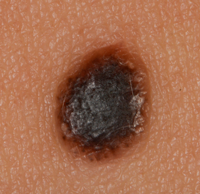
A new change to an area of skin (lesion) that was abnormal and turned out to be melanoma
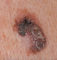
Doctors sometimes use the term lesion to describe a finding on the skin. This means an area of skin that looks different from the surrounding area.
Melanoma on the back
The following 2 examples contain 2 pictures. The first picture in each example is taken from a distance. The second picture from each example is a close up of the melanoma.
Example 1 - Picture of a melanoma from a new, dark lesion on the skin
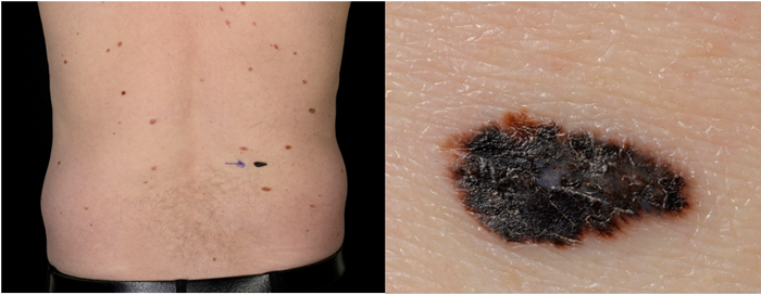
Example 2 - Picture of melanoma that may or may not have developed from a mole

Melanoma that has developed from a changing area of the skin with an irregular shape and colour
The blue markings in this picture below outline the area where the melanoma is. This is to show the surgeon the area they need to remove.
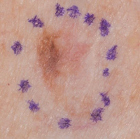
Melanoma that hasn't developed from a mole and is starting to spread
